
For pathologists and laboratories
Run the test and issue results on site
Digistain Breast runs in a clinical laboratory, on surgical resection tissue the laboratory already holds.
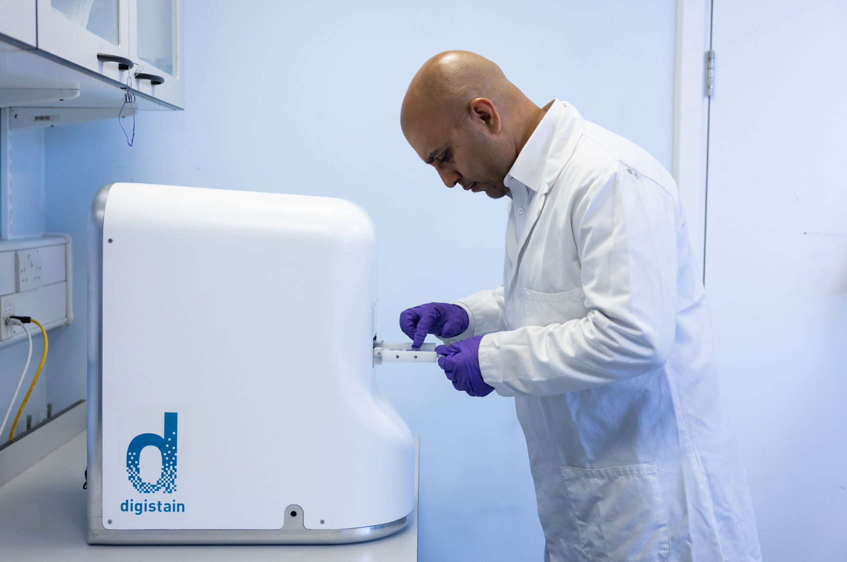
Running a Digistain analysis in the laboratory.
Workflow
How a laboratory runs Digistain Breast
Select the specimen
Surgical resection FFPE tissue. Core biopsies are not suitable.
Cut sections
Unstained sections. [NUMBER AND THICKNESS TO CONFIRM AGAINST THE IFU]
Acquire spectra
Mid-infrared measurement on the released instrument configuration.
Issue the report
Digistain Breast software produces the prognostic score. [REPORT FORMAT TO CONFIRM AGAINST THE IFU]
Specimen requirements
What the test needs
- Surgical resection FFPE sections only.
- Core biopsies are excluded.
- Patients: HR-positive (ER+ and/or PR+), HER2-negative, stage I–II invasive breast cancer, 0–3 positive lymph nodes, treated with adjuvant endocrine therapy alone.
- In-scope and out-of-scope specimen list: [TO CONFIRM]
Released configuration
Instrument
Thermo Fisher Nicolet iN10, at a fixed 400 × 400 µm aperture.
For use by trained laboratory personnel in a clinical laboratory.
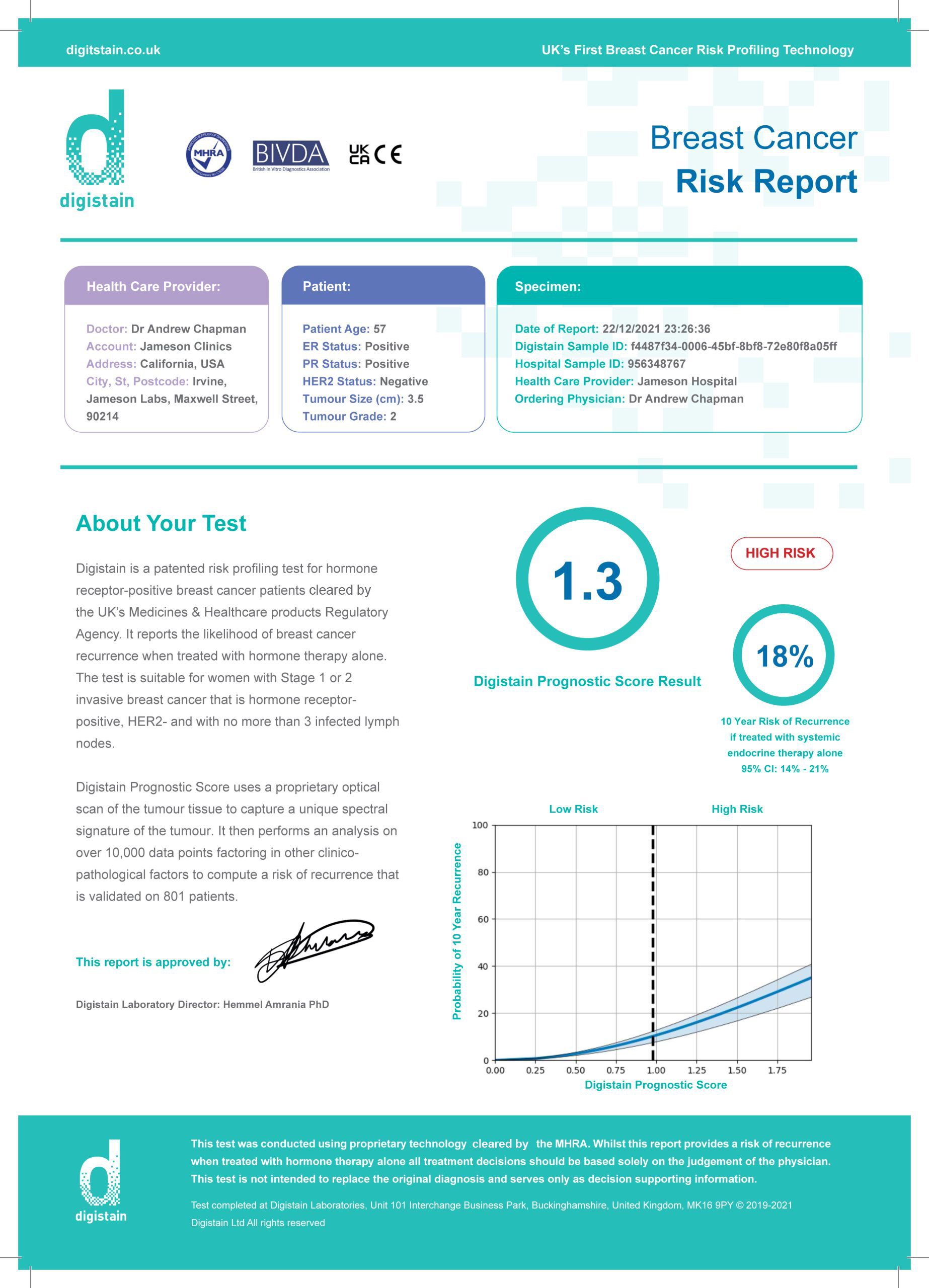
Report
Example report
An example of the report the laboratory issues. [REPORT FORMAT TO CONFIRM AGAINST THE IFU]
Training and support
Supported from the first sample
Digistain trains laboratory staff on the test, specimen requirements and reporting. [TRAINING AND SUPPORT PACKAGE TO CONFIRM]
Alternative route
Use Digistain’s laboratory service
Laboratories that do not run the test can send samples to Digistain’s laboratory. Usually three to four days including shipping, depending on courier and customs.
Laboratories
Bring Digistain Breast into your laboratory
Talk to us about the workflow, the released configuration and training.
Regulatory status and intended use
Intended use. Digistain Breast is in vitro diagnostic software that gives a quantitative prognostic score for risk of recurrence and recurrence-free survival at 5 and 10 years. It is used as a categorical (High/Low) prognostic indicator alongside other clinicopathological factors in pre- and post-menopausal women with HR-positive (ER+ and/or PR+), HER2-negative, stage I–II invasive breast cancer with 0–3 positive lymph nodes, treated with adjuvant endocrine therapy alone. It is not intended for diagnosis, for predicting response to therapy or for selecting therapy, and it is not a companion diagnostic. For use by trained laboratory personnel in a clinical laboratory, on surgical resection FFPE sections. Core biopsies are not suitable.
United Kingdom: UKCA-marked (self-declared), CE-marked under the IVDD, and registered with the MHRA.
European Union: CE-marked under the IVDD. IVDR Class C conformity assessment in progress with notified body QMD Services (2962).
United States: Not currently available in the US.
Other markets: [STATUS PER MARKET]
ISO 13485:2016 certified (LRQA) for the design and development of IVD software used in cancer management.